Klinefelter Syndrome

Klinefelter Syndrome

Klinefelter syndrome is characterized by small, firm testes, azoospermia (no sperm count), gynecomastia, in men with two or more X chromosomes. The common karyotype is 47,XXY . They have elevated levels of plasma gonadotropins. They have male psychosexual orientation and function sexually as normal men.

It is the most frequent major abnormality of sexual differentiation. This condition exists in roughly 1 out of 1000 males.  1 in 500 males have an extra chromosome but may not have the syndrome.

The syndrome was named after Dr. Harry Klinefelter, who worked with Fuller Albright and first described it in the year 1942.

The principal effects are development of small testicles and reduced fertility. 

  • Before puberty the testes appear normal but are small.
  • Gynecomastia (enlargement of breasts ) develops during adolescence, is generally bilateral and painless.
  • Mean body weight is increased due to longer legs.
  • Obesity and varicose veins occur in one-third to one-half.
  • Mild mental deficiency, social maladjustment.
  • Abnormal of thyroid functions, diabetes mellitus, and pulmonary disease may be present.
  • The risk of breast cancer is 20 times that of normal men (but only about a fifth that in women)

Treatment : Gynecomastia may need surgical treatment. Testosterone injections are needed.

Turner Syndrome

Turner Syndrome
This condition is a disorder of sexual development of women. It arises due to defective sexual differentiation at the time of fertilization. Turner syndrome is a genetic condition in which a female does not have the usual pair of two X chromosomes.

Humans have 46 chromosomes. Two of these chromosomes, the sex chromosomes, determine if you become a boy or a girl. Females normally have two of the same sex chromosomes, written as XX. Males have an X and a Y chromosome (written as XY).

In Turner syndrome, cells are missing all or part of an X chromosome. The condition only occurs in females. Most commonly, the female patient has only one X chromosome. Others may have two X chromosomes, but one of them is incomplete. Sometimes, a female has some cells with two X chromosomes, but other cells have only one.

About half of individuals with Turner syndrome have monosomy X, which means each cell in the individual’s body has only one copy of the X chromosome instead of the usual two sex chromosomes.Turner syndrome can also occur if one of the sex chromosomes is partially missing or rearranged rather than completely absent.Some women with Turner syndrome have a chromosomal change in only some of their cells, which is known as mosaicism. These are called mosaic Turner syndrome.Which all genes on the X chromosome are responsible for most of the features of Turner syndrome is still not fully understood. One gene called SHOX that is important for bone development and growth, and missing one copy of this gene is a likely causes for short stature and skeletal abnormalities in women with Turner syndrome.Most cases of Turner syndrome are not inherited. When this condition results from monosomy X, the chromosomal abnormality occurs as a random event during the formation of reproductive cells (eggs and sperm). An error in cell division called nondisjunction can result in reproductive cells with an abnormal number of chromosomes. For example, an egg or sperm cell may lose a sex chromosome as a result of nondisjunction. If one of these atypical reproductive cells contributes to the genetic makeup of a child, the child will have a single X chromosome in each cell and will be missing the other sex chromosome.Mosaic Turner syndrome occurs as a random event during cell division in early fetal development. As a result, some of an affected person’s cells have the usual two sex chromosomes (either two X chromosomes or one X chromosome and one Y chromosome), and other cells have only one copy of the X chromosome.
The incidence is estimated at 1 in 3000 newborn females.

Main features in this condition are absence of menstruation (Primary amenorrhea), incomplete development of sexual characters (sexual infantilism), short stature, multiple congenital abnormalities in women. After the time of expected puberty pubic and axillary hair remain sparse, the breasts are infantile, and no menses occur. In a small percentage of cases some menstruation may occur. Indeed, occasionally minimally affected women become pregnant; the reproductive life in such individuals is brief.

Main features:

  • Absence of menstruation. In this condition the egg cells in the ovaries usually die prematurely and most ovarian tissue degenerates before birth. A majority of the girls do not undergo puberty unless they are treated with the hormone estrogen. A small percentage of females with Turner syndrome do retain normal ovarian function.
  • After the time of expected puberty pubic and axillary hair remain sparse, the breasts are infantile, and no menses occur. In a small percentage of cases some menstruation may occur. Indeed, occasionally minimally affected women become pregnant; the reproductive life in such individuals is brief.
  • Incomplete development of sexual characters resulting to sexual infantilism.
  • Short stature, becomes evident at about the age of 5.
  • Multiple congenital abnormalities in women.
  • Webbed Neck is seen in about 30 percent of people. This is due to extra folds of skin on the neck.
  •  A low hairline at the back of the neck.
  • Drooping of the eyelids
  • Ears are differently shaped. Ears are set lower on the sides of the head than usual.
  • Abnormal bone development especially the bones of the hands and elbows.
  • A larger than usual number of moles on the skin.
  • Puffiness or swelling (lymphedema) of the hands and feet.
  • Congenital heat diseases: coarctation of the aorta, the aortic valve defect.
  • Some girls with the condition may experience learning difficulties, particularly in math. Many have a difficult time with tasks that require skills such as map reading or visual organization. Many women with Turner syndrome have normal intelligence and some may have higher than average intelligence.

People with TS are all different. Some may have many physical abnormalities and symptoms, whereas others experience only a few medical problems. With early and appropriate medical care and ongoing support, many with TS can lead normal, healthy, and productive lives.

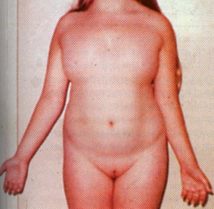
The diagnosis is made either at birth because of the associated anomalies or at puberty when amenorrhea and failure of sexual development are noted.The external genitalia are of female type but remain immature and do not grow as in adult females. There is no breast development. Internally the fallopian tubes and uterus are also immature. On the both sides the ovaries are grossly underdeveloped.Skeleton and the connective tissue are also involved. Swelling of the hands and feet, webbing of the neck, low hairline, skin folds on the back of the neck, a shield like chest with widely spaced nipples, and growth retardation. These features suggest the diagnosis in infancy. Ears may be deformed.  A fishlike mouth, Eyes may show ptosis – drooping of the upper eye lids.

Other associated conditions which may be present in such patients are : Coarctation of Aorta, Renal malformations, tendency to keloid formation, hearing impairment, unexplained hypertension, tendency to diabetes, thyroid disease.
Treatment : At the anticipated time of puberty, replacement therapy with estrogen should be started. This helps in the maturation of the breasts, labia, vagina, uterus, and fallopian tubes. Linear growth and bone maturation rates are also improved by this treatment. But the eventual height  rarely approaches the predicted level. Combination therapy with oxandrolone and/or growth hormone accelerates growth and increases final height.
 Genetic Disorders

Haemophilia

Haemophilia A

Haemophilia A is a genetic disorder characterised by defficiency of Factor VIII in the blood. The factor VIII gene is located on the X chromosome . Thus this disorder is a sex linked disorder.

  • One in 10,000 males is born with deficiency or dysfunction of the factor VIII molecule.
  • Although normal hemostasis requires at least 25 percent factor VIII activity, symptomatic patients usually have factor VIII levels below 5 percent.
  • Patients with <1 percent factor VIII activity have severe disease.
  • Patients with levels between 1 and 5 percent have moderatedisease with less frequent bleeding episodes.
  • Those with levels over 5 percent have mild disease with infrequent bleeding that is usually secondary to trauma.
  • Occasional patients with factor VIII levels as high as 25 percent are discovered when they bleed after major trauma or surgery.
  • The majority of patients with Haemophilia A have factor VIII below 5 %
  • All daughters of haemophilics are carriers and sisters have a 50 % chance of being a carrier.
  • If a carrier has a son he has 50 % chance of having haemophilia, and a daughter has 50 % chance of of being a carrier.
  • Haemophilia ‘breeds true’ within a family. All members of a family will have same abnormality of factor VIII gene, i.e. if one member has severe form of the disease all other affected would have severe form of the disease.
  • In carrriers of this disease, Ratio of factor VIIIC : von Willibrand factor (vWF) is reduced compared to the normal.
  • Tracing of the haemophilia gene within families can be done using gene probes which detect restriction fragment length polymorphism (RFLPs).
  • Antenatal diagnosis can be done in females having high degree of probability of being a carrier. This is done by doing chorion villus sampling at 8-9 weeks of gestation.

Haemophilia B

Hemophilia B is similar to Hemophilia A. Both are abnormality of blood coagulation. Hemophilia B is caused by the deficiency of Factor IX. It is also known as Christmas disease. It occurs in 1 in 100,000 male births. It has to be diagnosed accurately because clinically it is indistinguishable from factor VIII deficiency – Hemophilia A.

Factor IX is one of a group of six proteins synthesized in the liver that require vitamin K for biologic activity.

Treatment of Haemophilia A

Plasma products enriched in factor VIII have revolutionized the care of hemophilia patients, reduced the degree of orthopedic deformity, and permitted virtually any form of elective and emergency surgery. Now this mode of treatment is being widely used.

The widespread use of factor VIII concentrates also has produced some serious complications, including viral hepatitis, chronic liver disease, and AIDS.

Cryoprecipitate, which contains about half the factor VIII activity of fresh frozen plasma in one-tenth the original volume, is simple to prepare and is produced in hospital or regional blood banks. It must be stored frozen and is thawed and pooled before administration. Partially purified factor VIII concentrate, which is prepared from multiple donors and supplied as a lyophilized powder, can be refrigerated and reconstituted just before use.

There had been immense problem with the safety of these products. Three major developments have increased the safety of factor VIII therapy. First, heating of lyophilized factor VIII concentrates under carefully controlled conditions can inactivate human immunodeficiency virus (HIV) without destroying factor VIII coagulant activity. Second, highly purified factor VIII can be produced by adsorbing and eluting factor VIII from monoclonal antibody columns. Third, recombinant factor VIII is now available.

Patients with hemophilia should receive either monoclonal purified or recombinant factor VIII to minimize viral infections and other complications.

It has been determined empirically that each unit of factor VIII infused, defined as the amount present in 1 ml normal plasma, will raise the plasma level of the recipient by 2 percent per kilogram of body weight. Factor VIII has a half-life of 8 to 12 h, making it necessary to infuse it continuously or at least twice daily to sustain a chosen factor VIII level. In patients with mild hemophilia, an alternative to the use of plasma products is desmopressin (DDAVP), which transiently increases the factor VIII level. Desmopressin, in general, will increase the factor level two- to threefold. Although generally safe, it occasionally causes hyponatremia or may precipitate thrombosis in elderly patients.

  • An uncomplicated episode of soft tissue bleeding or an early bleeding into a joint can be treated with one infusion of sufficient factor VIII concentrate to raise the factor VIII level to 15 or 20 percent. A more extensive  bleeding requires twice-daily or continuous infusions in order to keep the factor VIII level between 25 and 50 percent for at least 72 h. Life-threatening bleeding into the central nervous system or major surgery may require therapy for 2 weeks with levels kept at a minimum of 50 percent of normal.
  • Patients with joint involvement also need skilled orthopedic care with immobilization of inflamed joints to promote healing and to prevent contractures and physical therapy to strengthen muscles and maintain joint mobility.
  • Hemophiliacs also require treatment before dental procedures. Filling of a carious tooth can be managed by a single infusion of cryoprecipitate or factor VIII concentrate coupled with the administration of 4 to 6 g of e-aminocaproic acid (EACA) four times daily for 72 to 96 h after the dental procedure. EACA is a potent antifibrinolytic agent that will inhibit plasminogen activators present in oral secretions and stabilize clot formation in oral tissue.

Most hemophiliac patients have had multiple episodes of hepatitis, and a majority have elevated hepatocellular enzyme levels and abnormalities on liver biopsy. Between 10 and 20 percent of patients also have hepatosplenomegaly, and a small number develop chronic active or persistent hepatitis or cirrhosis. A few patients with hemophilia have received liver transplants with cure of both diseases.

Following multiple transfusions, between 10 and 20 percent of patients with severe hemophilia develop inhibitors to factor VIII. Inhibitors are, generally, IgG antibodies that rapidly neutralize factor VIII activity. Before surgery, every patient should be screened for the presence of an inhibitor to factor VIII.

Female carriers of hemophilia, who are heterozygotes, usually produce sufficient factor VIII from the factor VIII allele on their normal X chromosome for normal hemostasis. However, occasional hemophilia carriers will have factor VIII levels far below 50 percent due to random inactivation of normal X chromosomes in tissue producing factor VIII. These symptomatic carriers may bleed with major surgery or bleed occasionally with menses. Rarely, true female hemophiliacs arise from consanguinity within families with hemophilia or from concomitant Turner’s syndrome or XO mosaicism in a carrier female.

Huntington's chorea

Huntington’s chorea

Chorea, the Greek word for “dance,” is used to describe the involuntary movements of the body especially of arms, legs and face. In Huntington’s chorea, these movements tend to be spastic and ballistic, and they dramatically affect normal functioning.

Huntington’s chorea is a dominantly inherited disease which is passed down through families by an autosomal dominant form of inheritance. It was first described by George Huntington in 1872. Incidence is 5-10/100,000.

The disease often presents as “nervousness“, and depression. Eventually progresses to include dementia and slowed eye movements. In juvenile HD, rigidity and parkinsonian tremor may be the primary manifestation. Generally, symptoms are first observed when the victim is in the ’20s or ’30s, and the degeneration slowly progresses, ending in the victim’s death some 10 to 20 years later.

The disease progresses with increasing chorea and loss of mental abilities. Eventually, abnormal movements and dementia become so severe that the person can no longer care for his or her self. 

Huntington’s chorea is due to slow degeneration in the basal ganglia, which eventually leads to cell death in the brain and the decrease and increase of various neurotransmitters. The symptoms of the disease are caused by a significant reduction (volume and activity) of two principal neurotransmitters (naturally occurring chemicals in the brain) – namely Acetylcholine and GABA, in turn affecting the activity of the neurotransmitter Dopamine, which becomes hyperactive. The disorder is partly characterized by an increase in the availability of dopamine, which can cause symptoms of chorea. HD is a basal ganglia disease; the portions most severely affected are caudate and putamen.

Huntingtons can be diagnosed on MRI by caudate atrophy with appropriate history, and also by genetic testing.

At present, there is no cure. The aim is to treat symptoms and support and protect the patient. Drugs can control the chorea somewhat, but benefits are often outweighed by the side effects. Caretakers must see to the person’s basic needs.

These include

  • hygiene
  • skin care
  • bowel and bladder care
  • feeding.

One must stay alert for suicide attempts. The person’s family often needs emotional support. Caring for the person at home is often beyond the family’s capacity.

Down syndrome

Down syndrome

It is one of the most frequently occurring chromosomal abnormalities found in humans, occurring once in approximately every 800 to 1,000 live births.
John Langdon Down, an English physician, published an accurate description of a person with Down syndrome. It was this scholarly work, published in 1866, which earned Down the recognition as the “father” of the syndrome. Although other people had previously recognized the characteristics of the syndrome, it was Down who described the condition as a distinct and separate entity.
In 1959, the French physician, Jerome Lejeune, identified Down syndrome as a chromosomal anomaly. Instead of the usual 46 chromosomes present in each cell, Lejeune observed 47 in the cells of individuals with Down syndrome.

Down syndrome affects people of all races and economic levels. Women age 35 and older have a significantly increased risk of having a child with Down syndrome. A 35-year-old woman has a one in 400 chance of conceiving a child with Down syndrome, and this chance increases gradually to one in 110 by age 40. At age 45 the incidence becomes approximately one in 35.

Diagnosis

The diagnosis of Down syndrome is usually suspected after birth as a result of the baby’s appearance.

There are many physical characteristics which form the basis for suspecting an infant has Down syndrome. If Down syndrome is suspected, a karyotype will be performed to ascertain the diagnosis. Some infants with Down syndrome have only a few of these traits, while others have many.

Among the most common traits are:

  • Muscle hypotonia, low muscle tone
  • Flat facial profile, a somewhat depressed nasal bridge and a small nose.
  • Oblique palpebral fissures, an upward slant to the eyes.
    Dysplastic ear, an abnormal shape of the ear. Simian crease, a single deep crease across the centre of the palm.
  • Hyper flexibility, an excessive ability to extend the joints
    Dysplastic middle phalanx of the fifth finger, fifth finger has one flexion furrow instead of two.
  • Epicanthal folds, small skin folds on the inner corner of the eyes.
  • Excessive space between large and second toe.
  • Enlargement of tongue in relationship to size of mouth.

All people with Down syndrome have some level of mental retardation. however, the level usually falls into the mild to moderate range .Children with Down syndrome learn to sit, walk, talk, play, toilet train and do most other activities, only somewhat later than others. Speech is often delayed.

Most people with Down syndrome have IQs that fall in the mild to moderate range of retardation. Children with Down syndrome are definitely educable

Today those with Down syndrome are working in various jobs and doing productive work. Banks, hotels, music, computer and many types of industries are employing individuals with Down syndrome.

People with Down syndrome date, socialize and form on-going relationships. Some are beginning to marry. Women with Down syndrome can and do have children, but there is a 50 percent chance that their child will have Down syndrome. Men with Down syndrome are believed to be sterile, with only one documented instance of a male with Down syndrome who has fathered a child.

Children with Down syndrome are at increased risk for certain health problems. Congenital heart defects, increased susceptibility to infection, respiratory problems, obstructed digestive tracts and childhood leukemia occur with greater frequency among children who have Down syndrome. However, advances in medicine have rendered most of these health problems treatable, and the majority of people born with Down syndrome today have a life expectancy of approximately fifty-five years.

Adults with Down syndrome are at increased risk for Alzheimer’s disease. Whereas approximately 6% of the general population will develop the disease, the figure is about 25% for people with Down syndrome.

Up to 50 percent of individuals with Down syndrome are born with congenital heart defects. The majority of heart defects in children with Down syndrome can now be surgically corrected with resulting long-term health improvements.

Leukemia – Individuals with Down syndrome have a 15 to 20 times greater risk of developing leukemia. The majority of cases are categorized as acute megakaryoblastic leukemia, which tends to occur in the first three years of life, and for which there is a high cure rate. A transient form of leukemia is also seen in newborns with Down syndrome, disappearing spontaneously during the first two to three months of life.

Screening tests for Down syndrome

There are two types of procedures available to pregnant women: Screening tests and diagnostic tests. Screening tests estimate the risk of the fetus having Down syndrome; diagnostic tests tell whether or not the fetus actually has the condition.

The most commonly used screening tests are the Triple Screen and the Alpha-fetoprotein Plus. These tests measure quantities of various substances in the blood (alpha-fetoprotein, human chorionic gonadotropin and unconjugated estriol) and together with the woman’s age, estimate her risk of having a child with Down syndrome. These screening tests are typically offered between fifteen and twenty weeks of gestation.

Screening tests are of limited value and are often performed in conjunction with a detailed sonogram. These tests are only able to accurately detect about sixty percent of fetuses with Down syndrome.

The procedures available for prenatal diagnosis of Down syndrome are chorionic villus sampling (CVS), amniocentesis and percutaneous umbilical blood sampling (PUBS). Each one of these procedures carries a small risk of miscarriage as tissue is extracted from the placenta or the umbilical cord to examine the fetus’s chromosomes. The procedures are about 98 to 99 percent accurate in the detection of Down syndrome.