Thalassaemia

Thalassaemia

Thalassaemia is inherent impairment of haemoglobin production due to partial or complete failure to synthesize a specific type of globin chain.

Hereditary disorders that result in a structurally abnormal hemoglobin or an insufficient quantity of hemoglobin are the most common genetic diseases of humans.

The production of hemoglobin requires a supply of iron, synthesis of heme, and synthesis of globin. Hemoglobin is essential for normal oxygen delivery by red blood cells.

When the condition is Heterozygous, the production of hemoglobin is mildly affected and the condition causes little disability. The synthesis is grossly affected when the patient is Homozygous.

In thalassaemias there is decrease in the quantity of normal globin chains in the hemoglobin. Thalassaemias are divided into the alpha thalassaemias, in which it is the production of alpha globin is deficient, and the beta thalassaemias, in which beta globin production is defective. The thalassaemias are divided clinically into Thalassaemia Minor and Thalassaemia Major, based on disease severity.

Beta-Thalassaemia

In this there is failure to synthesize beta globin chains. It is the most common type and is seen in highest frequency in the Mediterranean area. Those who are heterozygous, are thalassaemia minor, the condition is mild with mild anaemia and usually no disability. The homozygous are thalassaemia major, have profound anaemia after first 4 months of life.

Beta Thalassaemia major
Causes profound anaemia which has crippling effect on the health of the child. The child needs blood transfusion and chances for survival without transfusion for few years are low. Changes in the bone marrow (hyperplasia), results in head bossing and prominent malar eminences, which gives typical appearance of these diseases.

Development and growth of the child is retarded.
Folic acid deficiency develops.
Spleenomegaly (enlargement of spleen) is an early feature.
Cardiac enlargement with heart failure is common.

Repeated transfusions give rise to haemosiderosis (excessive deposition of iron ).

Beta Thalassaemia minor
Is detected often after treatment for mild anaemia with iron therapy fails. The condition is mild and often without any symptom.

Management of Beta Thalassaemia

  • Blood transfusion to maintain haemoglobin.
  • Bone marrow transplantation – Allogeneic bone marrow transplantation from HLA complatible sibling.
  • Folic acid 5 mg daily.
  • In case of iron overload – Desferrioxamine therapy.
  • Splenectomy performed as late as possible.

Prevention
During early in pregnancy, DNA analysis of chorionic villus material can be done to identify a fetus with homozygous beta thalassaemia. Such pregnancy can be terminated. This examination can be done if both the parents are known to be carriers (thalassaemia minor).

Alpah Thalassaemia

It is due to reduction or absence of alpha chain synthesis. This type is common in the South East Asia. There are 2 alpha gene loci on chromosome 16 are therefore four alpha genes. If one is deleted there is no clinical effect. If two are deleted there may be a mild hypochromic anaemia. If three are deleted the patient has Hemoglobin H disease. If all four are deleted the baby is still born. Treatment of Hemoglobin H is similar to beta thalassaemia of intermediate severity.

Genetic Disorders

Klinefelter Syndrome

Klinefelter Syndrome

Klinefelter syndrome is characterized by small, firm testes, azoospermia (no sperm count), gynecomastia, in men with two or more X chromosomes. The common karyotype is 47,XXY . They have elevated levels of plasma gonadotropins. They have male psychosexual orientation and function sexually as normal men.

It is the most frequent major abnormality of sexual differentiation. This condition exists in roughly 1 out of 1000 males.  1 in 500 males have an extra chromosome but may not have the syndrome.

The syndrome was named after Dr. Harry Klinefelter, who worked with Fuller Albright and first described it in the year 1942.

The principal effects are development of small testicles and reduced fertility. 

  • Before puberty the testes appear normal but are small.
  • Gynecomastia (enlargement of breasts ) develops during adolescence, is generally bilateral and painless.
  • Mean body weight is increased due to longer legs.
  • Obesity and varicose veins occur in one-third to one-half.
  • Mild mental deficiency, social maladjustment.
  • Abnormal of thyroid functions, diabetes mellitus, and pulmonary disease may be present.
  • The risk of breast cancer is 20 times that of normal men (but only about a fifth that in women)

Treatment : Gynecomastia may need surgical treatment. Testosterone injections are needed.

Turner Syndrome

Turner Syndrome
This condition is a disorder of sexual development of women. It arises due to defective sexual differentiation at the time of fertilization. Turner syndrome is a genetic condition in which a female does not have the usual pair of two X chromosomes.

Humans have 46 chromosomes. Two of these chromosomes, the sex chromosomes, determine if you become a boy or a girl. Females normally have two of the same sex chromosomes, written as XX. Males have an X and a Y chromosome (written as XY).

In Turner syndrome, cells are missing all or part of an X chromosome. The condition only occurs in females. Most commonly, the female patient has only one X chromosome. Others may have two X chromosomes, but one of them is incomplete. Sometimes, a female has some cells with two X chromosomes, but other cells have only one.

About half of individuals with Turner syndrome have monosomy X, which means each cell in the individual’s body has only one copy of the X chromosome instead of the usual two sex chromosomes.Turner syndrome can also occur if one of the sex chromosomes is partially missing or rearranged rather than completely absent.Some women with Turner syndrome have a chromosomal change in only some of their cells, which is known as mosaicism. These are called mosaic Turner syndrome.Which all genes on the X chromosome are responsible for most of the features of Turner syndrome is still not fully understood. One gene called SHOX that is important for bone development and growth, and missing one copy of this gene is a likely causes for short stature and skeletal abnormalities in women with Turner syndrome.Most cases of Turner syndrome are not inherited. When this condition results from monosomy X, the chromosomal abnormality occurs as a random event during the formation of reproductive cells (eggs and sperm). An error in cell division called nondisjunction can result in reproductive cells with an abnormal number of chromosomes. For example, an egg or sperm cell may lose a sex chromosome as a result of nondisjunction. If one of these atypical reproductive cells contributes to the genetic makeup of a child, the child will have a single X chromosome in each cell and will be missing the other sex chromosome.Mosaic Turner syndrome occurs as a random event during cell division in early fetal development. As a result, some of an affected person’s cells have the usual two sex chromosomes (either two X chromosomes or one X chromosome and one Y chromosome), and other cells have only one copy of the X chromosome.
The incidence is estimated at 1 in 3000 newborn females.

Main features in this condition are absence of menstruation (Primary amenorrhea), incomplete development of sexual characters (sexual infantilism), short stature, multiple congenital abnormalities in women. After the time of expected puberty pubic and axillary hair remain sparse, the breasts are infantile, and no menses occur. In a small percentage of cases some menstruation may occur. Indeed, occasionally minimally affected women become pregnant; the reproductive life in such individuals is brief.

Main features:

  • Absence of menstruation. In this condition the egg cells in the ovaries usually die prematurely and most ovarian tissue degenerates before birth. A majority of the girls do not undergo puberty unless they are treated with the hormone estrogen. A small percentage of females with Turner syndrome do retain normal ovarian function.
  • After the time of expected puberty pubic and axillary hair remain sparse, the breasts are infantile, and no menses occur. In a small percentage of cases some menstruation may occur. Indeed, occasionally minimally affected women become pregnant; the reproductive life in such individuals is brief.
  • Incomplete development of sexual characters resulting to sexual infantilism.
  • Short stature, becomes evident at about the age of 5.
  • Multiple congenital abnormalities in women.
  • Webbed Neck is seen in about 30 percent of people. This is due to extra folds of skin on the neck.
  •  A low hairline at the back of the neck.
  • Drooping of the eyelids
  • Ears are differently shaped. Ears are set lower on the sides of the head than usual.
  • Abnormal bone development especially the bones of the hands and elbows.
  • A larger than usual number of moles on the skin.
  • Puffiness or swelling (lymphedema) of the hands and feet.
  • Congenital heat diseases: coarctation of the aorta, the aortic valve defect.
  • Some girls with the condition may experience learning difficulties, particularly in math. Many have a difficult time with tasks that require skills such as map reading or visual organization. Many women with Turner syndrome have normal intelligence and some may have higher than average intelligence.

People with TS are all different. Some may have many physical abnormalities and symptoms, whereas others experience only a few medical problems. With early and appropriate medical care and ongoing support, many with TS can lead normal, healthy, and productive lives.

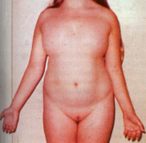
The diagnosis is made either at birth because of the associated anomalies or at puberty when amenorrhea and failure of sexual development are noted.The external genitalia are of female type but remain immature and do not grow as in adult females. There is no breast development. Internally the fallopian tubes and uterus are also immature. On the both sides the ovaries are grossly underdeveloped.Skeleton and the connective tissue are also involved. Swelling of the hands and feet, webbing of the neck, low hairline, skin folds on the back of the neck, a shield like chest with widely spaced nipples, and growth retardation. These features suggest the diagnosis in infancy. Ears may be deformed.  A fishlike mouth, Eyes may show ptosis – drooping of the upper eye lids.

Other associated conditions which may be present in such patients are : Coarctation of Aorta, Renal malformations, tendency to keloid formation, hearing impairment, unexplained hypertension, tendency to diabetes, thyroid disease.
Treatment : At the anticipated time of puberty, replacement therapy with estrogen should be started. This helps in the maturation of the breasts, labia, vagina, uterus, and fallopian tubes. Linear growth and bone maturation rates are also improved by this treatment. But the eventual height  rarely approaches the predicted level. Combination therapy with oxandrolone and/or growth hormone accelerates growth and increases final height.
 Genetic Disorders

Homocystinuria

Homocystinuria

It is an inborn error of Methionine metabolism in which there is deficiency of enzyme cystathionine b-synthase

Homocysteine is converted to methionine. In homocystinuria there is impaired conversion of homocysteine to methionine, The sulfur atom of the essential amino acid methionine is transferred ultimately to cysteine by the transsulfuration pathway. In one of these steps, homocysteine condenses with serine to form cystathionine. This reaction is catalyzed by the pyridoxal phosphate-dependent enzyme cystathionine b-synthase.

Homocysteine and methionine accumulate in cells and body fluids; cysteine synthesis is impaired, resulting in reduced concentrations of this amino acid and its disulfide form cystine.

Homocystinuria is relatively common in Ireland (1 in 60,000 births) but rare elsewhere (less than 1 in 200,000 births).

This genetic defect is also seen in consanguineous marriages.

Features:
Mental retardation
Venous thromboses- a major cause of morbidity
Osteoporosis
Hair often fine & brittle. Malar flush. Ectopia lentis, myopia.
Convulsions in about 10%. Psychiatric disturbances.
Skeletal features resembling Marfan syndrome – in 50 % of cases.

Diagnosis:
Detection of Homocysteine in urine.

Range of Homocysteine.in blood

  • Normal ( Male) : – 8.0 – 14.0 mmol/L
  • Normal (Female) : – 6.0 -12.0 mmol/L
  • Moderate homocystenaemia : – 16-30 mmol/L
  • Intermediate homocystenaemia : – 31-100 mmol/L
  • Severe homocystenaemia : – > 100 mmol/L

Treatment:
Pyridoxine 20-300 mg per day.

Restricted intake of methionine plus cystine supplements.
Administration of choline or betaine to enhance remethylation of homocystine

Homocysteine is an amino acid that’s normally produced in the body in small amounts from the amino acid methionine. Homocysteine’s role in the body is to control growth, and support bone and tissue formation.
Scientists have known that people with high concentrations of the amino acid homocysteine in the blood are at a much higher risk for stroke and heart disease. When homocysteine levels rise, they begin to damage arteries and stimulate growth of arteriosclerotic plaques which leads to heart disease.

High levels of homocysteine can be caused by

  • Vitamin deficiencies
  • Normal aging
  • Thyroid problems
  • Kidney disease
  • Genetic disorders like Homocystinuria.

Study shows folic acid and vitamins B6 and B12 can decrease homocysteine concentrations in the blood – reducing the risk of heart disease.

Anyone who has a high risk for heart disease either from family history or poor nutritional background, or who has early signs of heart disease probably should take vitamin supplements to control their homocysteine level and stop the arteriosclerotic process.

Severe deficiency of Folic Acid, vitamin B6 and B12 in the elderly may result in elevated serum homocysteine levels. Its a new risk factor for the Cardiac disease. Deficiency of each one of these vitamins may lead to accumulation of homocysteine which is known to have toxic effect on the vascular tissue.

A study found high levels of homocysteine in second generation Indian immigrants as compared to Europeans.

Folic acid rich foods are – Pulses, dried beans, green leafy vegetables.

Pyridoxine ~ Vitamin B6

Malaria

Malaria

Malaria is a type of fever which is a major cause of morbidity in the developing countries. Despite world-wide attempts to eradicate this disease it continues to be a big problem.
Caused by bite by female Anopheles mosquito which trasmits four types of parasites of genus Plasmodium – vivax, ovale, falciparum, malariae.
Plasmodium vivax is the most common and the Plasmodium falciparum is the most deadly.
Malaria is charactirised by relapsing fever with shaking chills, prostration, anaemia, and enlarged spleen.
Fever paroxysms are characterised by three phases:
Cold phase – lasting for 1-2 hours, during which sudden feeling of cold is followed by shivering, then intense rigors.
Hot phase – lasting for 3-4 hours, when there are hot flushes with headache and exhaustion as temperature reaches its peak.
Wet phase – lasting for 2-4 hours, when profuse sweating occurs as temperature returns to normal.
Fever paroxysms occur
every 48 hours in case of P.vivax, P.ovale,
every 72 hours in case of P. malariae.
In case of P.falciparum periodic fever paroxysms are not seen rather irregular intermittent fever or daily paroxysms may be seen.

Other symptoms – nausea, vomiting, cough, joint pains, abdominal and joint pain. Pallor and jaundice.

Death due to vivax malaria is very rare. Most malaria related fatalities occur due to P. falciparum malaria that can kill a non immune person within a week or two of infection.

Red Blood Cells which are invaded by the P.falciparum become very sticky and attach themselves to the walls of the capillaries. This blocks the microcirculation and thus causes cellular hypoxia, hypoglycemia, lacticacidosis and increased cellular permeability. These changes result in cerebral, pulmonary and renal manifestations.

Complications of Falciparum Malaria.
1. Cerebral Malaria – should be suspected in cases with history of short fever followed by deep unconsciousness in endemic areas. This accounts for 80% of deaths from acute malaria.
2. Algid Malaria – characterised by vomiting, nausea, diarrhoea, dehydration, low blood pressure, rapid respiration and low urine output.
3. Blackwater Fever – is caused by sudden extensive destruction of RBCs in the blood. Haemoglobin is found in the urine and this condition may precipitate renal failure. At times this condition is precipitated by the administration of Quinine to the patient.
4. Pernicious Anaemia – when blood contains a very high number of malaria parasite leading to rapid destruction of the RBCs.
5. Pulmonary Oedema and Adult Respiratory distress syndrome.
6. Vascular Collapse and Shock with Hypothermia and Adrenal Insufficiency.
7. Febrile Convulsions.
8. Hyperpyrexia
9. Metabolic Acidosis

In the management of Malaria the important and critical aspect is the diagnosis and treatment of falciparum malaria as soon as possible as deterioration of the patient’s condition can be rapid and sudden.
Besides drug treatment management of serious sign and symptoms are also very important.

Drug treatment of Malaria
Chloroquine: 600 mg immediately, 300 mg after 6 hrs., 300 mg on day 2, 300 mg on day 3 — for all four types of malaria.

Artemether: For Chloroquine resistant falciparum malaria.
day 1 – 80 mg IM twice.
Next 4 days 80 mg Once daily.

Quinine: For Chloroquine resistant falciparum malaria.
Slow infusion over 4 hrs. Initial loading dose 20 mg /kg followed every 8 hrs. with 10mg / kg. for 10 days.

Mefloquine: For Chloroquine resistant falciparum malaria.
750 mg immediately. 500 mg 8 hrs. later.

Primaquine: given after a course of chloroquine for the elimination of P. vivax and P. ovale from the body.

Other drugs: Pyrimethamine/sulfadoxine combination. Doxycycline.

Caution :
Quinine or Chloroquine not to be given as IV bolus.
Dexamethasone IV , Manitol, Heparine to be avoided in Cerebral Malaria

Prophylactic medication for Malaria.

In endemic areas regular intake of chloroquine 2 tabs. for adults or Pyrimethamine/sulfadoxine 1 tab. for adults weekly may be use to prevent malaria.